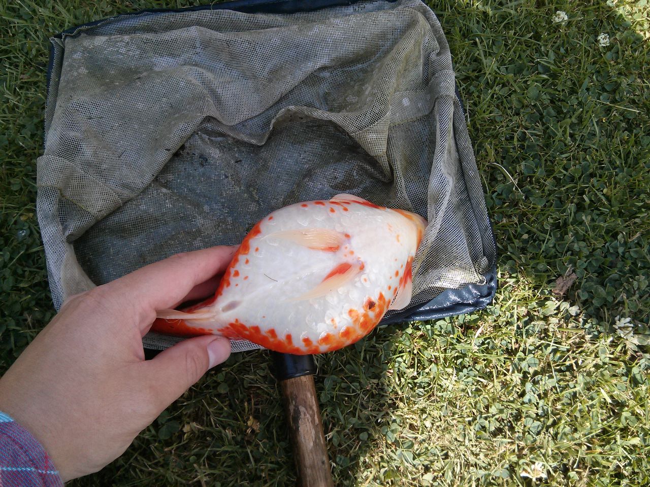
Bloated Belly: What to Do About Dropsy?

This is a common but difficult problem and typifies the problems involved in fish disease diagnosis. Abdominal swelling is not a disease – but a clinical sign of several possible health problems. Because an internal disorder is involved, in most cases it isn’t possible to say what the problem was until a post mortem is carried out. The most common causes are:
- A genetic disorder that usually shows as the fish grows older. In the early stages this seems to cause little discomfort for the fish and it will lead a normal life for some time – often several years.
- A tumour or growth. The only option in this situation is surgery, which clearly requires professional help. However, the survival rate of such procedures is very low as this is still very much an experimental procedure.
- Systemic bacterial infection, which is usually accompanied by raised scales, protruding eyeballs and sometimes reddening / inflammation on the body. If caught early enough this may respond to a course of antibiotic injections. Bath treatments are rarely successful
- Viral diseases: Much the same signs as bacterial infections but no cure
- Internal organ disease – such as heart problems- leading to an accumulation of fluid in the abdominal cavity. This leads to a balloon-shaped swelling and the abdomen feels very soft and sqidgy – unlike a tumour which tends to feel hard. No cures, and heart transplants are just not on.
- Intestinal blockage / constipation: This is more usually associated with loss of equilibrium, but in some severe cases it can lead to swelling. The only possible treatment is either try to feed the fish a few frozen peas, which act as a laxative, or else try baths in Epsom salts (70g / litre for 5 minutes) which has the same effect. If the condition is advanced, the success rate is likely to be poor
- Could indicate intestinal parasites. Making wet mounts of faeces for microscopic examination may assist diagnosis.